From single cell to human fetus: a detailed look at your baby’s first three months
First Trimester Development
- Your pregnancy countdown starts from the beginning of your last menstrual period.
- At just five weeks, your pregnancy will be visible on ultrasound.
- By six weeks, the regular flicker of a heartbeat is first visible on ultrasound.
- At nine weeks, your baby's head is large in relation to the trunk, and the arms and legs are short and flipper-like.
- By the end of the first trimester, your baby's body proportions have changed to become similar to those of a newborn.
 fter first discovering they’re pregnant, most women can’t wait to find out more about the changes that are about to take place in their bodies. The first trimester is the most amazing three-month period in biology; what begins as a single cell (the fertilized egg) will become a tiny fetus with all of the baby’s internal organs in place and recognizable human features in just 13 short weeks. Here’s a look at your baby’s development in the first trimester:
Weeks 1 to 4
Your pregnancy countdown starts from the first day of your last menstrual period. About two weeks before you notice a missed period, an egg is released from your ovary and a sperm fertilizes it to create a zygote. This zygote divides repeatedly as it travels along the fallopian tube. By the time it reaches the uterus, it's a bundle of 12 to 15 cells.
While the uterus waits for its arrival, it transforms itself into a welcoming home, ready to accept and nurture the zygote when it appears. The uterus creates extra blood vessels and special glands in its inner lining to nourish the developing baby. Four to five days after fertilization, the zygote has implanted itself in the uterus, where cell division and growth continue throughout your pregnancy.
Week 5
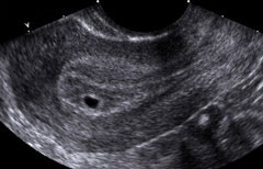
About one week after your missed period, your pregnancy will be visible on ultrasound. At this point, it is a small fluid collection called a gestational sac. It is just 2/10 of an inch in diameter and lies within the lining of the uterus known as the deciduas. (See Below.) Even though the baby is still too small to see, this first visual proof of the developing pregnancy is very exciting.
|
5-week ultrasound | 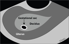 |
Week 6
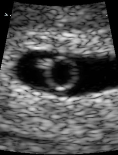
A week later, the baby is finally visible on ultrasound. (See Below.) At this point, it appears as a tiny oval blip approximately a tenth of an inch in length (about the width of a sunflower seed). The regular flicker of a heartbeat is first visible on the screen.
| 6 week ultrasound | 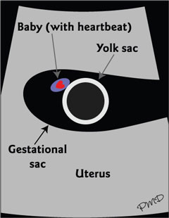 |
Weeks 7 to 10
The most striking change over the next two to three weeks is the appearance of identifiable human features. The baby now has recognizable parts, such as head, body, arms and legs. The umbilical cord is also present. Although human features are present, the proportion is much different than it will be when your baby is born. The head is large in relation to the trunk, and the arms and legs are short and flipper-like. At nine weeks, your baby will be 9/10 of an inch in length–about the size of a grape. The baby is also quite active at this stage, but you won't be able to feel any movements for at least 10 more weeks.
| 9 week 3-D ultrasound |

Weeks 11 to 13
By the end of the first trimester, the baby's body proportions have changed to become much more similar to those of a newborn. By 13 weeks, the baby is three inches long, measured from the top of the head to the bottom of the rump. (About the size of a small peach.) This represents a 30-fold increase in length in the seven-week interval from six to 13 weeks gestation.
|
13-week 3-D Ultrasound |  |


0 comments:
Post a Comment